Funding

Three active grants build one machine: an MRI scanner rebuilt from its gradient and receiver chains outward. Thirty years of funded work sit behind them.
3Active grants
52Completed
1994First grant
2029Runs through
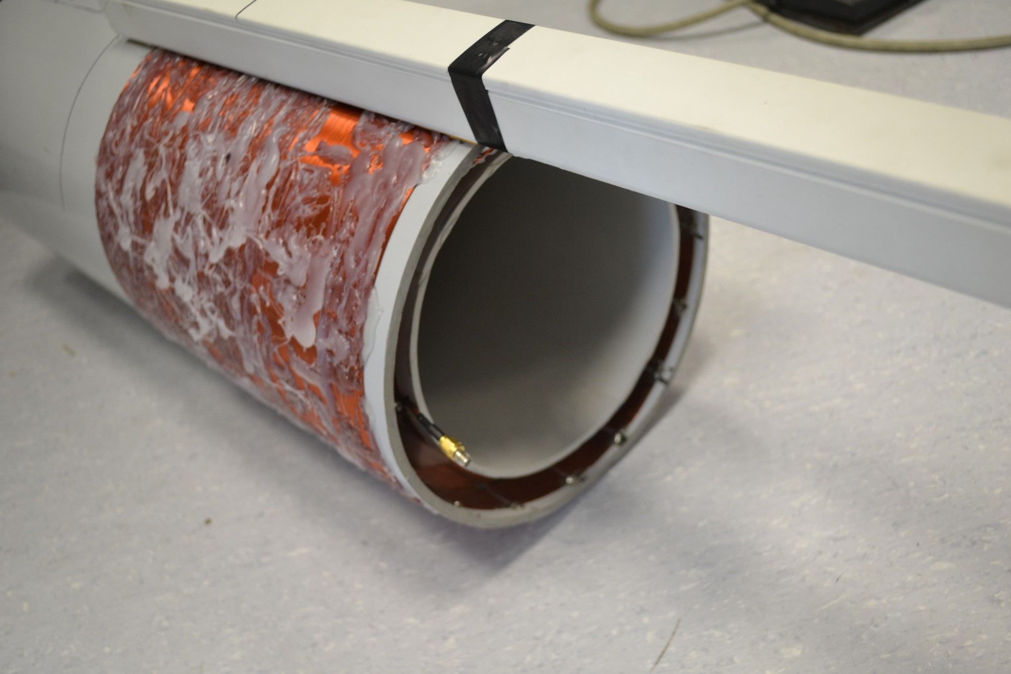
Gradient array coil wound in copper, built in the Ergin Atalar Lab

Active

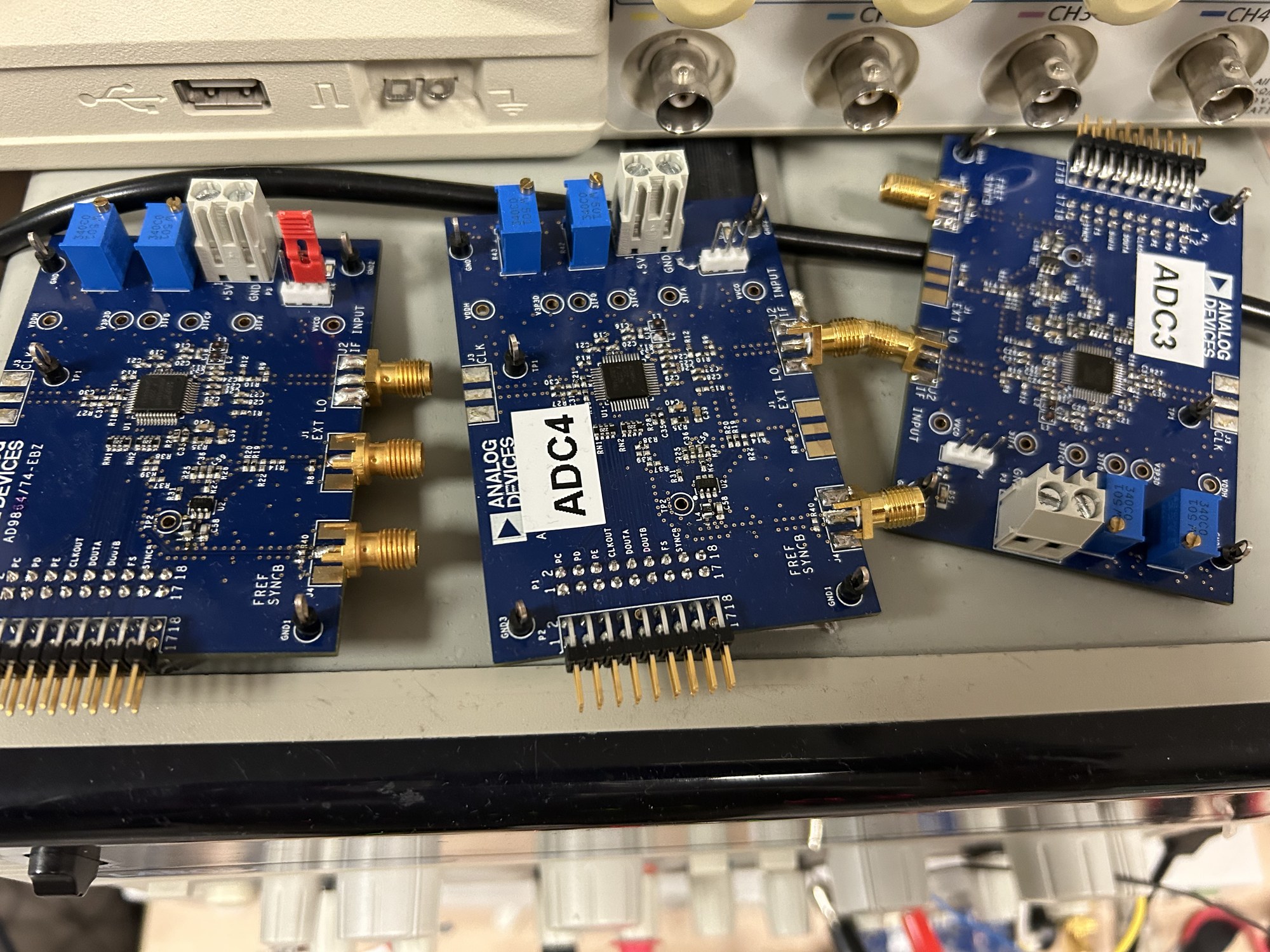
Evaluation boards of the receiver converter chip, one per channel
Converter boards on the bench, one per channel.
2023–2027TÜBİTAK 100423AG005

High Performance Digital Receiver for MRI with Optical Communication

Yüksek performanslı optik iletişimli sayısal MR alıcısı geliştirilmesi

Principal Investigator, Project 10Leader of the Bilkent component
The signal is digitized where it is picked up, and leaves the magnet as light.
Part of a platform project. İnsan Fonksiyonunu Tehdit Eden Zorluklara Karşı Nöroteknolojik Çözümler Platformu, under the Mükemmeliyet Merkezi Destek Programı. Coordinator: Can Yücesoy. The Bilkent University component funds three subprojects, led by Tolga Çukur, Emine Sarıtaş and Ergin Atalar.

A conventional MRI scanner carries the receive signal out of the magnet room as an analog voltage on a coaxial cable. The cable collects noise on the way. It also couples to the transmit field, so every channel needs decoupling, and the cable bundle sets a practical ceiling on the number of channels.

In this subproject we digitize the signal where it is picked up. An ultra-low-noise amplifier, a filter and a mixer sit next to the coil element, and the converter follows them on the same board. We build the chain for both proton frequencies, 64 MHz at 1.5 T and 123 MHz at 3 T. What leaves the element is light on an optical fibre, not a voltage on copper. The same fibre carries power, data and the clock the other way, so no copper crosses the bore.

The difficulty is in the budget. The converter has to run on the power an optical link can deliver, which is why the design carries a voltage booster and an energy store. The clock has to be recovered from that link and stay stable enough for imaging, which is why it carries a temperature-controlled crystal oscillator. A microcontroller supervises the whole element.

The result is a receiver that scales. Adding a channel means adding a fibre. It no longer means another coaxial cable and another decoupling network.
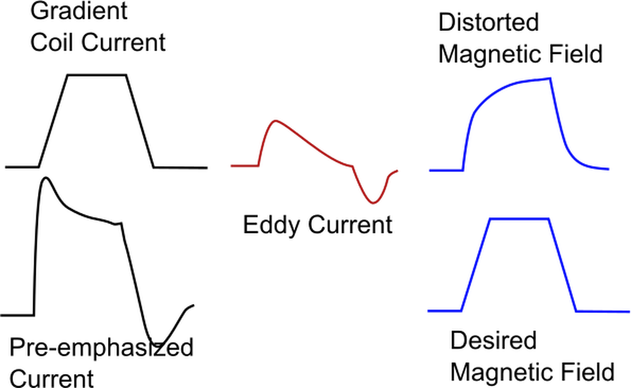
Pre-emphasised coil current, the induced eddy current, and the resulting magnetic field
Pre-emphasis: the current is distorted so the field is not.
2026–2029TÜBİTAK 1001125E832

Eddy Current Compensation with Gradient Arrays in Magnetic Resonance Imaging

Manyetik Rezonans Görüntülemede Gradyan Dizileri için Girdap Akım Telafisi

Principal Investigator
The emerging gradient array technology enables the acquisition of images that cannot be obtained with conventional MRI systems.

In Magnetic Resonance Imaging (MRI), the gradient magnetic fields used can induce eddy currents in the magnet that produces the static magnetic field, leading to two significant undesirable effects. First, the eddy currents can heat the superconducting magnet, potentially disrupting its superconductivity. This can result in high repair costs and render the imaging system inoperable for several days. In addition to this critical issue, the magnetic fields generated by eddy currents can distort the resulting images. These well-known problems have established mitigation strategies in conventional MRI systems.

The emerging gradient array technology enables the acquisition of images that cannot be obtained with conventional MRI systems. However, while the eddy current problem has established solutions in conventional systems, it remains an unresolved issue for systems utilizing gradient arrays.

In this project, we aim to develop innovative methods that reduce eddy currents and correct the resulting magnetic field distortions when using gradient array technology. In our preliminary work, we developed solution approaches that required extensive computation time. The proposed method provided a more effective solution to the eddy current problem than conventional systems. However, due to reliance on a commercial finite element software, these methods proved impractical for real-world application.

In the first work package of the project, we plan to achieve significantly faster solutions for electromagnetic calculations by using magnetoquasistatic methods. In the second work package, we will represent the frequency-domain calculations as a multi-input multi-output (MIMO) filter. In the final work package of the project, we will demonstrate the use of our developed method to compensate for eddy currents induced by fundamental imaging sequences when using a gradient array system that we are currently developing.

As a result of this project, we will eliminate yet another barrier to the practical implementation of a gradient array technology that has the potential to fundamentally transform magnetic resonance imaging techniques.
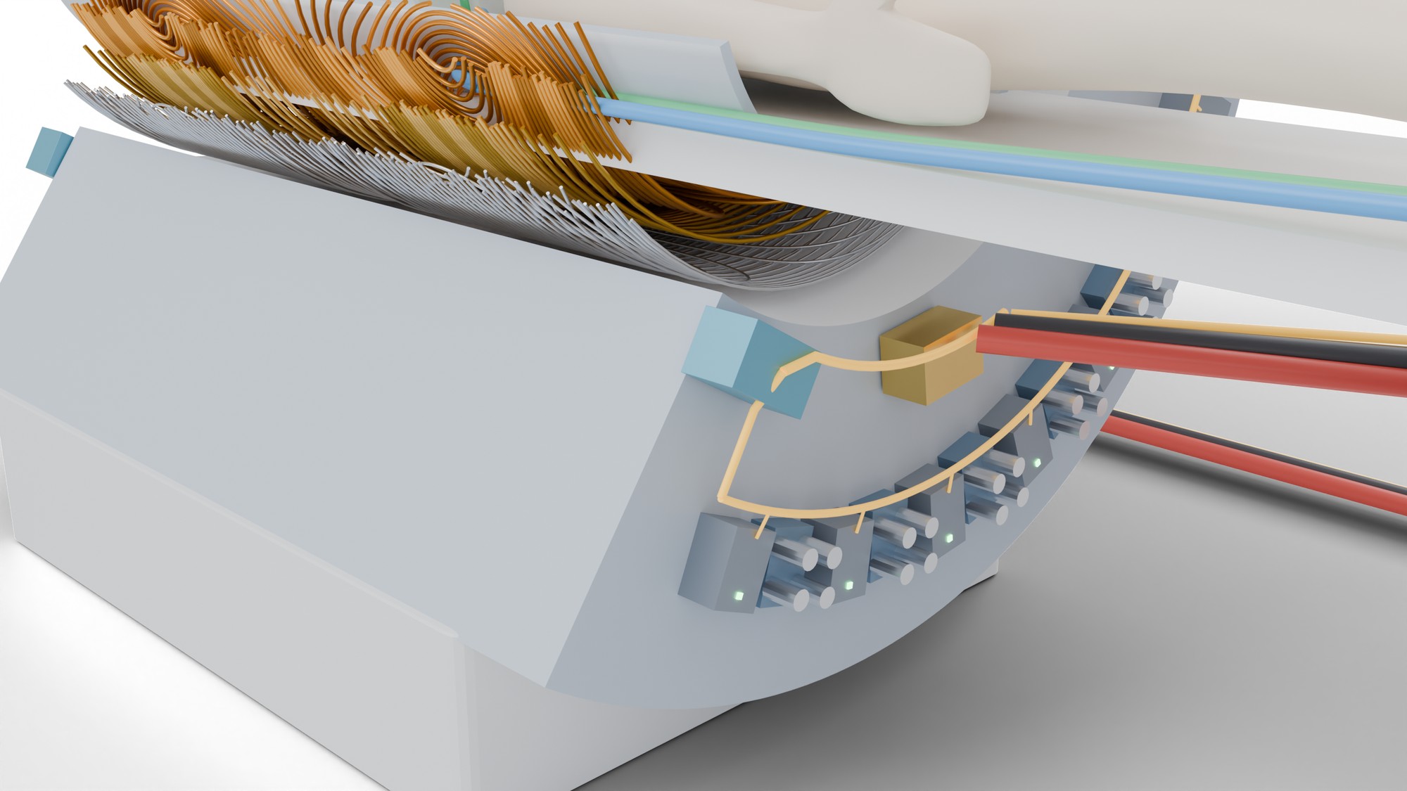
Illustration of a gradient array with an amplifier placed beside each coil element
Illustration: an amplifier beside every coil element.
2025–2027TÜBİTAK 2247-B125C001

A Massive Gradient Array for Magnetic Resonance Imaging: More is Different

ERC Projeleri Güçlendirme Desteği Programı

Principal Investigator
The gradient systems are the engines of magnetic resonance imaging (MRI) scanners.

The gradient systems are the engines of magnetic resonance imaging (MRI) scanners. The conventional MRI scanners are equipped with three coil-amplifier pairs. Expensive and very powerful amplifiers drive the x-, y- and z-gradient coils. The switching speed and strength of the gradient field determine the image quality and speed, but the nerve stimulation limits these due to the induced electric field in the body. The distribution of the magnetic field and the body’s position determine the electric field. In conventional systems, linear magnetic fields are generated in a large diameter of a spherical volume (DSV) independent of how patients are located inside the scanner. However, linear fields are not necessary in a large DSV at all times during imaging. The changing need for linearity cannot be obtained with conventional design.

The project proposes a design for a massive gradient array system that maximizes the nerve stimulation threshold dynamically by modulating the electric field distribution in the body while providing the needed magnetic field distribution during imaging. The proposed design will also enable imaging in the conventional imaging mode with the large DSV to ensure compatibility with the existing technologies. Unlike the conventional systems, many inexpensive digitally controlled amplifiers will be in the close vicinity of the coil elements. Complete digital control of the system will enable maximum utilization of the available hardware, provide high fidelity gradient fields and generate the highest quality brain images.

Currently, MRI is the most powerful diagnostic imaging instrument. The wealth of diagnostic information is limited by available imaging time and cost. The gradient hardware limitations hold many exciting imaging techniques for accurately diagnosing devastating diseases. The proposed massive gradient system will eliminate one of the critical barriers to further developing MRI technology.

The aim of this TÜBİTAK 2247-B project is to collect preliminary data to strengthen the ERC Advanced Grant application.

Completed  ·  fifty-two grants, 1994–2025

2020s
2023–2025
TUBITAK 1001 123E024Manyetik Rezonans Görüntüleme Cihazları Için Gradyan Dizisi Ve Rf Verici Dizisi Sürme Yetenekli Spektrometrenin Tasarımı Ve Gerçekleştirilmesi
Principal Investigator
2021–2023
TUBITAK 1001 121E128Design and Implementation of a Set of Z-Gradient Array and an Active-Shield Array for Magnetic Resonance Imaging
Co-Investigator
2021–2023
TUBITAK JPCOFUND 121N028 The EU Joint Programme – Neurodegenerative Disease Research (JPND)Spinocerebellar ataxias: Advanced imaging with ultra-high field MRI
Principal Investigator of Work Package 5
2010s
2019–2022
TUBITAK 1001 119E116Manyetik Rezonans Goruntuleme Cihazlari icin Dairesel Radyo-Frekans Sargi Dizilerinin Analizi, Tasarimi ve Yapimi
Principal Investigator
2019–2021
ASELSANMR Goruntuleme Sistemi Gelistirme Projesi
Principal Investigator
2018–2021
TUBITAK 1001 117E817Beyin Manyetik Rezonans Elastografisinin Guvenligi ve Optimizasyonu
Principal Investigator
2017–2020
Strateji ve Butce BaskanligiPreklinik Goruntuleme Yaridimi ile Asi Test ve Gelistirme
Principal Investigator
2017–2022
ASELSAN, (supported by TUBITAK TEYDEB)Cok Kanalli Gradyan Bobin Dizisi Tasarimi, Sinyal Ureteci ve Kontrol Karti Gelistirilmesi ve Prototip Uretimi
Principal Investigator
2017–2018
TUBITAK 1000Bilkent Universitesi Ar-Ge Strateji Belgesi (Sinir Bilimleri)
Principal Investigator
2017–2020
TUBITAK 1001 (116E270)Manyetik Rezonans Goruntuleme Cihazi İçin Ozellestirilmis E-Sinifi Sayisal Radyo Frekans Yukselteci Dizisi
Principal Investigator
2015–2016
ASELSAN36 Kanalli RF Bobin Dizisi Tasarimi, Gelistirilmesi ve Prototip Uretimi
Principal Investigator
2014–2016
SANTEZImplant Uyumlu MR Cihazi (Implant Compatible MR Scanner)
Principal Investigator
2014–2017
TUBITAK 1001 (114E186)Etkin Ayristirma Yontemi Kullanarak Sifir Eko Zamanli Manyetik Rezonans Goruntulemesi (Zero Echo Time Magnetic Resonance Imaging Using Active Decoupling Technique)
Principal Investigator
2013–2015
TR Ministry of DevelopmentUlusal Manyetik Rezonans Arastirma Merkezi Faz II (National Magnetic Resonance Research Center (UMRAM) Phase II)
Principal Investigator
2013–2016
TUBITAK 1001 (103S959)Ailesel El Titremesi (Essential Tremor-ETM)’nin Genetik, Yapisal ve Islevsel Temellerinin Arastirilmasi
Consultant
2013–2016
TUBITAK 1001 (102K069)Insan Beyninin Yapisal Ve Islevsel Mimarisinin Genetik Kokenli Merkezi Sinir Sistemi Bozukluklari Incelenerek Arastirilmasi
Consultant
2013–2016
TUBITAK 3501 (113E187)Manyetik Rezonans Goruntuleme Altinda Hizli Ve Guvenilir Ozgul Sogurma Orani Ve Isi lerinin Gelistirimesi
Consultant
2013–2016
TUBITAK 3501 (114E167)Kanser Goruntuleme Icin Manyetik Parcacik Goruntuleme: Bolgesel Difuzyon Algilama
Consultant
2013–2016
TUBITAK 3501 (114E546)Kategori Hedefli Dogal Görsel Taramanin Insan Beyninde Görsel Temsillere ve Islevsel Baglantilara Etkisi
Consultant
2013–2016
TUBITAK 3501 (113S001)Dejeneratif Resesif Ataksilerin Nozolojisi ve Molekoler Tanisi
Co-Investigator
2012–2016
TUBITAK 1001 (112E555)Dogrusal Olmayan Graydanlar Manyetik Alanin Manyetik Rezonans Goruntulemesindeki Uygulamalari (Applications of Nonlinear Gradient Fields in Magnetic Resonance Imaging)
Principal Investigator
2010–2013
TUBITAK 1001 (109E226)Verici Sargi Dizilimi Ile Manyetik Rezonans Goruntulemesi Sirasinda Olusan Ozgul Sogurma Hizinin Azaltilmasi
Principal Investigator
2010–2013
TUBITAK TEYDEBEndoservikal Manyetik Rezonans Goruntuleme
Principal Investigator
2010–2013
TUBITAK 1001Femtosaniye Nanobiyofotonik icin Ileri Fiber Cozumleri
Consultant
2000s
2009–2010
University of Minnesota, A Collaborative ProjectProducing Miniaturized RF Signal Fiber Optic Transmission System for 7 Tesla MRI Scanners
Principal Investigator
2009–2012
TUBITAK 1001Baglamin Gorsel Algiya Etkileriyle Ilgili Olarak Insan Korteksindeki Sinirsel Etkinliklerin ve Islevsel Baglantilarin Incelenmesi
Consultant
2009–2012
TUBITAK 1001Insanlarda Gelisimsel Sinir Sistemi Hastaliklarina Neden Olan Mutasyonlarin Gorsel ve Motor Sisteminin Yapisal ve Islevsel Organizasyonuna Etkilerinin Arastirilmasi
Consultant
2008–2011
TUBITAK 1001Girisimsel MRG de Kateterlerin Yerinin Otomatik Olarak Belirlenebilmesi Icin Yeni Bir Tumlesik Mikro Optoelektronik Sistem
Consultant
2007–2011
SANTEZManyetik Rezonans Uyumlu Elektrofizyoloji Araclari
Principal Investigator
2007–2011
TUBITAK 1001Manyetik Rezonans Goruntulemesi Yonteminin Hastalara Takilan Uyarici Elektronik Cihazlar Uzerindeki Etkilerinin Incelenmesi
Principal Investigator
2007–2009
TUBITAK TEYDEBManyetik Rezonans Goruntuleme Uyumlu Elektrofizyoloji Araclari
Principal Investigator
2007–2009
DPTGirisimsel ve Tanisal Manyetik Rezonans Arastirma Merkezi
Principal Investigator
2004–2006
FP6 MRIG-CT-2004-506262Magnetic Resonance Imaging Compatible Cardiac Pacemakers and Implantable Cardioverter-Defibrilators
Principal Investigator
2003–2008
NIH R01Imaging Cardiac Regeneration with Mesenchyma Stem Cell
Co-Investigator
2002–2005
NIH R21Mesenchymal Stem Cell and Ventricular Remodeling
Co-Investigator
2001–2004
NIH R01Digital optical imaging of vascular gene expression
Co-Investigator
2001–2004
US ArmyEndo-urethral MRI guided Prostate Ablation
Principal Investigator
2001–2005
NIH R01Intravascular MRI-Enhanced Vascular Gene Transfer
Co-Investigator
2001–2006
NIH R01Innovative MRI Research Technology
Co-Investigator
2001–2004
NSF PERSupplement to Center for Medical Robotics and Computer-Assisted Interventional Systems and Technology
Principal Investigator
1990s
1999–2000
Gatewood FellowshipHigh Resolution Endouretral MRI of the Female Urethra and Urethral support tissues with patological correlation
Co-Investigator
1999–2001
CIRREFIVMRI guided vascular interventions
Co-Investigator
1999–2004
NIH R01Harmonic Phase MRI for Ultrafast Cardiac Strain Imaging
Co-Investigator
1998–1999
Whitaker Foundation, Biomedical Engineering Transitional GrantHigh Resolution Imaging and Spectroscopy of Atherosclerotic Plaques using Intravascular MRI
Principal Investigator
1998–2000
SurgiVision Inc., Research GrantDevelopment of MR Probes
Principal Investigator
1998–2003
NIH R01Intravascular Magnetic Resonance: Towards Clinical Interventions
Principal Investigator
1998–2008
NSF Engineering Research CenterCenter for Medical Robotics and Computer-Assisted Interventional Systems and Technology
Co-Investigator
1997–2005
NIH R01 HL057483MR-guided Coronary Balloon Angioplasty
Principal Investigator
1995–1998
Whitaker Foundation, Biomedical Engineering Research GrantHigh Resolution Imaging and Spectroscopy of Atherosclerotic Plaques using Intravascular MRI
Principal Investigator
1995–1999
NIH, SCOR is ischemic heart diseaseProject 6: Metabolic Characterization of Reperfused Dysfunction Myocardium by NMR
Co-Investigator
1995–2000
NIH R01Dynamic 3D tagged MRI of the Cardiac Cycle in Ischemia
Co-Investigator
1994–1996
US Army, Innovative developmental and exploratory awardIn-vivo microscopic MR imaging of breast lesions
Co-Principal Investigator